
Detection of avian influenza A viruses of subtype N5 by real-time RT PCR
This protocol is a copy of the standard operating procedure used by the WOAH/FAO International Reference laboratory for AI at the Animal and Plant Health Agency. If you have any technical queries please contact AIWRL@apha.gov.uk
WOAH/FAO international reference laboratory for AI
1. Introduction
1.1 Purpose and scope of this SOP
1.1.1 This protocol provides the details required to detect viruses of subtype N5 avian influenza virus (AIV) by reverse transcriptase RealTime (RRT)-
PCR in swabs, tissues and egg-amplified material.
1.2 Background information
1.2.1 Two versions of the N5 RRT-PCR are included in this protocol: (i) The N5 RRT-PCR assay amplifies specifically within a region of the neuraminidase (NA) gene of subtype N5 AIVs, with primers and probe designed for detection of N5 AIVs currently in global circulation, precisely as described by Hoffmann et al., 2016. (ii) The second version, referred to as the APHA-2016 N5 RRT-PCR (James et al., 2018), produces the same amplicon but the primers and probe sequences were modified for optimal detection of Eurasian H5N5 HPAIVs which emerged during 2016. Version (ii) is recommended for use in the first instance as the primers and probe set was designed to detect more-recently circulating HxN5 HPAIV strains than the primers and probe set from version (i). Furthermore, version (ii) is more user-friendly in employing two primers instead of four (James et al., 2019).
1.2.2 This protocol shares considerable commonality with other AIV RRT-PCRs in routine use at APHA Avian Virology (section 3), Procedure / Method (section 4) and Results (section 5) as regards (i) Chemicals and Reagents (3.2) for primers and probes, and (ii) the results interpretation guide for the two methods, i.e. sub-section 5.2.
2. Safety
2.1 It is your laboratory’s responsibility to ensure all work described in this protocol is conducted to a high safety standard.
3. Materials
3.1 Documentation and software
3.1.1 Procedures:
Procedure for isolation and detection of viruses from avian species using embryonated eggs.
RNA extraction from biological samples
Extraction of nucleic acids from swabs using the QIAGEN BioRobot Universal
Influenza-Detection of influenza A matrix gene by real time Taqman® RT-PCR
3.2 Chemicals and reagents
3.2.1 N5 RRT-PCR primers and probe, precisely as described by Hoffmann et al. (2016):
IAV-N5-1322F: 5'-AAG AGA GRA CWA GCA TTT GGA C-3'
IAV-N5-1353F:
5'-CTC CAC KGT RTT TTG TGG TGT-3'
IAV-N5-1421R:
5’-GGA AGA ATT GCK CCA TCA YC-3’
IAV-N5-1426R:
5’-CAA AKG GAA GAA TTG CKC CAT CA-3’
IAV-N5-1375FAM:
5'-FAM-TCM AGT GAG GTC CCA GGR TGG TC-BHQ1-3'
3.2.2 N5 real-time RRT-PCR primers and probe, APHA-2016 version :
N5 HR 16 1353F2:
5'-TTC CAC TGT GTT TTG TGG KGT-3'
N5 HR 16 1426R:
5’-CGA ATG GTA GAA TTG CTC CAT CA-3’
N5 HR 16 1375FAM2:
FAM-TCY AGT GAG GTC CCA GGA TGG TC-BHQ1
The probe is 5’ labelled with FAM and 3’ labelled with Black Hole Quencher (BHQ1).
3.2.3 Preparation of the N5 RRT-PCR master mix including the published primers and probes (see 3.2.1., Hoffmann et al., 2016) is described below. For the second version, simply substitute the APHA-2016 primers and probe (3.2.2.) which are provided as stock solutions at the same respective concentrations.
This utilises the Qiagen OneStep RT-PCR kit (Cat No. 1044287). Reagents not contained in the kit can be obtained from other suppliers.
Volumes indicated below will be sufficient for 10 x 25µl reactions, ie divide the master mix into 10 x 23µl volumes then add 2µl extracted RNA to each.
DEPC treated water (ie RNAse-free) | Ambion or similar | 130µl |
(x5) Qiagen 1 step RT PCR buffer | 50µl | |
Rox ref dye (pre-diluted 1:500 from stock in DEPC water) | Stratagene or similar | 3.75µl |
Qiagen dNTP mix | 10µl | |
IAV-N5-1322F: N5 (forward primer): (50µM) | Sigma or similar | 3µl |
IAV-N5-1353R: N5 (reverse primer): (50µM) | Sigma or similar | 3µl |
IAV-N5-1421R: N5 (reverse primer): (50µM) | Sigma or similar | 3µl |
IAV-N5-1426R: N5 (reverse primer): (50µM) | Sigma or similar | 3µl |
IAV-N5-1375FAM: N8 (30µM) Sigma or similar | Sigma or similar | 1.7µl |
25mM magnesium chloride | Promega or similar | 12.5µl |
RNAsin (40U/µl) | Promega or similar | 1µl |
Qiagen 1-step RT PCR enzyme mix | 10µl |
3.2.4 N5 RRT-PCR assay controls
1. Negatives:
Include at least one “no template controls” (NTC) where 2µl RNase-free water are added.
2. N5 AIV extraction control (positive):
The N5 RRT-PCR is never run in isolation (para 1.1.3) RNA extraction will be followed by up to three AIV RRT-PCR tests, namely M gene, H5 and H7, eg for IRL or Report Case submissions, or possibly at the early stage of an outbreak to help fully characterise the index case. Therefore the RNA extraction control may be an H5/H7 AIV isolate, and provided that this has given satisfactory results in the H5, H7 and / or M gene RRT-PCR, then there is no need to include an N5-positive extraction control.
3. N5 AIV RNA dilution series (“standards”):
Use an N5 AIV RNA preparation from extracted egg fluid recommended by the International Reference Laboratory as a 10-fold dilution series (to cover a range of at least 4 dilutions). This series (entered appropriately as “standards” with relative quantities indicated by using the software while setting up the RealTime experiment) can be used to determine the N5 RRT-PCR efficiency. See Appendix 1 for preparation of new batches
AIV RNA is prepared in batches with a designated arbitrary value of 107 and stored at -70C. Take out an aliquot and thaw. Carefully prepare ten-fold dilutions of RNA in DEPC water as follows:
Ten-fold dilution from 107 | Designated value (label tubes with these values) |
10-1 | 106 |
10-2 | 105 |
10-3 | 104 |
10-4 | 103 |
10-5 | 102 |
10-6 | 101 |
Mix each dilution by agitation / flicking and briefly centrifuge. Make sufficient of each dilution so that enough is made to use for all the N5 RRT-PCR runs for that day. Store the aliquots on ice. Discard at the end of the working day. Each standard to be run is indicated on the worksheet using the designated value.
If conducting semi-quantitative N5 AIV RRT-PCR, it is important to include low positive N5 AIV controls which correspond to 10 EID50/ml and 100 EID50/ml (i.e. approx. Ct 36) of N5 AIV in the sample prior to RNA extraction – this will relate to defining the N5 RRT-PCR positive / negative cut-off (section 5.2. below).
3.3 Equipment
3.3.1 Microcentrifuge tubes (1.5ml)
Biomek2000 robot or similar
-70ºC or lower freezer
20µl robot barrier tips
Pipettes
-18°C or lower freezer
Sterile, RNAse-free pipette tips with aerosol barrier
Vortex mixer
Mx3000/3005 or AriaMx plates/strips/caps
Microcentrifuge (with rotor for 2ml tubes)
Mx3000/3005 real time RT-PCR plate/strips/caps
Mx3000/3005 real time RT-PCR system
AriaMx RRT-PCR system
AriaMx real-time RT-PCR plates/strips/caps
AriaMx real-time PCR system
4. Procedure and method
4.1 Test reliability
4.1.1 Process of assuring ongoing test reliability:
Method & frequency: Internal QA (IQA) is achieved by blind testing in a Ring Trial. This consists of testing a “Mini Panel” of anonymous AIV samples, where IQA results demonstrate ongoing test reliability. In addition, an annual EU AI PCR Proficiency Panel is prepared & tested as part of an external QA (EQA) scheme, where testing of this Panel can also provide evidence of ongoing test reliability.
This and other AIV RRT-PCR protocols note the importance of gauging RRT-PCR performance by monitoring the Ct values and standard curve obtained from the 10-fold dilution series (RNA standards) to determine the efficiency of the RRT-PCR (see Appendix 1). This is crucial for the
routine use of the AI RRT-PCRs in Reference / Diagnostic Laboratory work.
4.2 Preparation of PCR master mix and loading
4.2.1 Preparation of PCR master mix and loading of RealTime plate / strips(s) to be carried out in a PCR clean room.
4.2.2 Make up master mix sufficient for the number of samples to be tested.
4.2.3 Thoroughly mix the master mix and centrifuge for 30 secs to remove bubbles.
4.2.4 Aliquot 23µl of master mix per well of the RealTime plate / strip(s).
Make sure the plasticware used is appropriate for the chosen RealTime PCR platform.
4.2.5 Loosely place the plate caps on the portion of the plate being used. All wells will need to be covered even though only a portion of the plate may be used.
4.2.6 Bring the plate / strip(s) out of the PCR clean room and place on ice before addition of sample RNA and controls.
4.3 Preparation of N5 RNA controls
4.3.1 N5 positive and negative controls are as described above in 3.2.4.
4.3.2 Extract subtype N5 AIV RNA manually or robotically.
4.3.3 Include subtype N5 AIV RNA positive controls “standards” as a dilution series in each N5 RRT-PCR experiment (section 3.2.4, paragraph 3).
4.4 Manual addition of samples and standards
4.4.1 NB Take care to avoid cross contamination of samples at this stage.
Change gloves frequently and do not hold tips containing RNA above the incorrect wells. It is advised to cover strips loosely which have not yet been filled with caps (strip covers) to further minimise the risk of cross contamination.
Referring to plate layout, add 2µl of the negative extraction control, then add the sample RNA (2µl) to the master mix. Replace caps on tubes.
Where possible, use a dedicated multi-channel pipette to load multiple samples e.g. eight samples per strip.
4.4.2 Add 2µl DEPC water as a no template control (NTC), replace cap on this well and then add the real time PCR RNA standards and positive RNA extraction control to appropriate wells based on your worksheet layout
4.4.3 Once RNA is added, fit caps (strip covers) to all wells.
4.4.4 It is important that the caps are fitted firmly and correctly onto the wells before insertion into the RealTime instrument.
4.4.5 If the RealTime plate/ strip(s) are not to be loaded into the instrument immediately, keep the plate / strip(s) on ice until ready to test.
4.5 Reverse transcription (RT) and PCR
4.5.1 Place the RealTime plate / strip(s) in the appropriate RealTime instrument. The instructions below are for the Agilent MX3000 PCR machine and indicate the settings that should be used.
4.5.2 The thermocycling profile is:
RT step:
45°C for 30 mins
95°C for 15 mins
PCR step (x40 cycles)
95°C for 15 secs
56°C for 20 secs
72°C for 30 secs
NB: These are different from our more commonly used H5 / H7 cycling conditions.
NB: Collect fluorescence data at end of 56°C step using the FAM and ROX filters. The HEX channel is chosen to detect VIC fluorescence. Use endpoint read option with one read.
N.B: ’Filter Gain Setting’ - After servicing or repair this setting is often changed back to the factory default of FAM x8. On return to APHA the MRM team routinely correct the filters for our use Cy5 ROX and HEX should all be set to x1, Fam should be set to x2. Please see example below. If prompted or you wish to check the filter settings, it is possible in the MXPro software to change the ‘Filter Set Gain Settings’ – click Instrument, then filter Set Gain Settings.
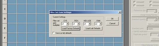
CY5 set to x1, ROX set to x1, HEXJDE set to x1, FAM set to x2
4.5.3 Open a new file on the instrument using the MxPro software and select the RealTime quantitative PCR (multiple standards) option for the experiment. The plate setup can be re-entered for each experiment or
imported from previous experiments if desired. The thermoprofile is imported from a dedicated folder stored at - S:\Virology\VI6 Research\MX
Data\1 Thermal profiles for import Note that the lamp on both RealTime machines requires 20 minutes to warm up, and this can occur during the RT step (4.5.2 above).
4.5.4 When the plate is setup and thermoprofile windows have been entered, select “run” to start thermocycling. A storage window will automatically open. By default the file should be saved in an appropriate folder on the S:/ drive on the APHA network, and ensure that the MxPro files are clearly named for N5 RRT-PCR.
4.5.5 The complete run takes approximately 2.0 hours. If the machine is not booked for immediate use by another colleague upon completion of the run, select the option to turn off the lamp at the end of the run. This is because the lamps have a limited lifespan.
5. Results
5.1 Analysis and display of results by using MxPro software
The analysis procedure described below is for the MX-Pro software and should be adapted for the software being used.
5.1.1 The fluorescence data can be viewed during and / or after the PCR using the raw data plots tab in the “Run” section by using the MxPro software.
5.1.2 When the run is complete, the MxPro file should be saved by default to an appropriate folder on the S:/ drive. Make a note of the file and folder names, see also 4.5.4.
5.1.3 To analyse the data select the Analysis section button and select the wells to be examined in the Analysis / Setup window.
5.1.4 To view the results click on the Results tab in the Analysis section and view the amplification plots. There are four options for analysing the fluorescence:
R (Multicomponent view) displays the raw fluorescence in arbitrary units.
dR displays the baseline-corrected fluorescence. As all reactions and wells will start with slightly different fluorescence readings, this option sets a baseline value of 0 to all plots. This correction is determined by the fluorescence values obtained during the initial rounds of the PCR. The adaptive baseline algorithm calculates the best baseline for each plot individually.
Rn displays the fluorescence normalised to the passive reference dye (ROX). This allows for fluctuations in fluorescence, which are not due to cleavage of the TaqMan probe.
dRn displays the baseline-corrected normalised fluorescence.
The dRn option with the ROX channel switched on is the most appropriate option for analysis of the data.
5.2 Interpretation of N5 RRT-PCR results
5.2.1 Analyse the data by comparing the results obtained for the negative (NTC) and N5 AIV positive controls.
5.2.2 Negative controls (NTCs):
All NTCs should give “No Ct” as their final result. High Cts in all NTC wells with a linear character (eg >37) & giving a very low level final fluorescence (ie little greater than the initial “flare” fluorescence values at early cycles) suggest that probe degradation may have occurred, eg the probe has been excessively frozen and thawed. Although such observations may not invalidate the experiment, it is advised to discard the aliquot & thaw-out a fresh aliquot of the relevant N5 probe for subsequent experiments & note result.
If the late Ct value (>37) has a logarithmic / sigmoidal character where clear final fluorescence values are observed, then contamination of the NTC wells with N8 AIV RNA may be considered. It is also possible that such very late Ct signals may occasionally occur spuriously. Repeat the N5 RRT-PCR experiment.
5.2.3 N5 AIV positive controls
1. N5 AIV extraction control (3.2.4, paragraph 2):
This should reproducibly amplify with a Ct value +/- 2 Cts either side of their predetermined Ct value. Greater increases above the predetermined Ct value suggest that:
RNA extraction from the specimens has been suboptimal. Repeat.
It is also possible that the N5 AIV positive control may have degraded if stored incorrectly eg 4C, excessive freeze-thawing etc….
Deviation (+/- 2 Ct values) may also occur where software settings on the instrument are inaccurate / inappropriate and the fluorescence cut-off level excessively high / low and is affecting the Ct value of the predetermined N5 AIV positive control. It may be possible to reset the software after the experiment to restore the expected Ct value provided the other controls are in order.
2. N5 AIV RNA dilution series “standards” (3.2.4, paragraph 3)
Ten-fold dilution series should yield a straight line with R2 value of >0.975 (ideally >0.985), slope in the range of -3.1 to –3.9 which should correspond to N5 RRT-PCR efficiency in the range 80 – 120%. Deviation from these conditions may be due to (i) inaccurate assembly of the master mix / inaccurate pipetting, (ii) inaccurate RNA addition (iii) degradation of the RNA standards by inappropriate storage or (iv) inefficient thermocycling / fluorescence reading due to an instrument problem. This indicates a need to address these matters for subsequent N5 RRT-PCR experiments.
5.2.4 If all controls are within acceptable limits, analyse the data for the test samples.
5.2.5 Criteria for assessing positive-negative results on test samples are as follows:
1. A test sample providing a Ct value < 36 in the N5 RRT-PCR is considered clearly N5 AIV-positive while a specimen recording “No Ct” value is clearly negative.
2. Test samples with Ct >36, but with a logarithmic / sigmoidal character giving clear final fluorescence greater than that observed during early cycles are generally considered as “inconclusive”. However, the following paragraph presents a possible strategy for confirmation as either “N5 [very low] positive” or “N5 negative”:
3. Check the Ct value when the same RNA extract was tested by the M gene RealTime PCR: A Ct value of 34-37 by M gene RealTime PCR suggests that this may be a very weak positive N5 RRT-PCR result. A reproducible result obtained after re-extraction of RNA from the same clinical material by N5 / M gene RealTime PCRs would tend to confirm this. However, it is unlikely that such “late Ct risers” (particularly those from field submissions) would yield positive results by other tests eg virus isolation or conventional AIV PCRs due to their lower sensitivity.
6. References
6.1 Hoffmann, B. et al. Riems influenza a typing array (RITA): An RT-qPCR based low density array for subtyping avian and mammalian influenza a viruses. Sci. Rep. 6, 27211; doi: 10.1038/srep27211 (2016).
http://www.nature.com/articles/srep27211
6.2 James. J., S.M. Reid, S. S. Thomas, S. Mahmood, A. M. P. Byrne, J. Cooper, C. Russell, B. C. Mollett, E. Agyeman-Dua, S. Essen, M. J. Slomka, I. H. Brown, and S. M. Brookes. Development and application of real-time PCR assays for specific detection of contemporary avian influenza virus subtypes N5, N6, N7, N8 and N9. 63(sp1), 209-218. doi:10.1637/11900-051518-Reg.1. (2019),
Appendix 1
Preparation of TaqMan®real-time RT- PCR RNA standards
These can be prepared from either live or inactivated egg-grown antigen and can be supplied either as an aliquot of freeze-dried material or an aliquot from a wet ‘antigen stock.
NB - For live antigen follow safety procedures in place for the virus type being produced. Work at the appropriate containment level for the virus being used – refer to the virus categorisation risk document
1. If the antigen is supplied freeze–dried, reconstitute the vial with 1ml molecular grade water. Ensure the solution is mixed thoroughly by gentle vortexing.
2. Manually extract the RNA from the whole reconstituted antigen from 1ml of wet stock according to the protocol for RNA extraction from biological samples.
3. Pool together the RNA extractions.
4. Freeze a 5µl aliquot at-70 ºC
5. Thaw this aliquot and prepare a 10-fold dilution series in molecular grade water from as described above from 10-1 - 10-7
6. Test the dilution series by the appropriate real-time RT-PCR. Test the current ‘in-use’ RNA standard batch in parallel on the same PCR test run.
7. Directly compare the results of the two batches. The Ct values and test efficiencies should be similar i.e. Ct values +/- 2.0 per corresponding dilution. If there is an apparent significant difference, adjust the dilution of the new batch of RNA (eg. make a 1/10 dilution) and repeat stages 3-7 above.
8. Once the required dilution is achieved, prepare 5µl aliquots, assign a batch number, label the tubes and store at -70 ºC.