
One Step real time RT-PCR for detection of H5 and H7 avian influenza virus and cleavage site sequencing
This protocol is a copy of the standard operating procedure used by the WOAH and FAO International Reference laboratory for avian influenza at the Animal and Plant Health Agency. If you have any technical queries, please contact AIWRL@apha.gov.uk
1. Introduction
1.1 Purpose and scope of this protocol
1.1.1 | This SOP provides details required to determine and analyse the nucleotide sequence of samples of H5 & H7 avian influenza (AI) submitted for statutory sequencing. PCR conditions outlined below include those which have been successful in generating amplicons from RNA directly extracted from clinical specimens. |
1.2 Background information
1.2.1 | As described in the OIE diagnostic manual, determination of the nucleotide sequence (and the deduced amino acid sequence) of specific regions of the genomes of these viruses allows an estimation of their pathogenicity to be made. |
2. Safety
2.1 | It is your laboratory’s responsibility to ensure all work described in this protocol is conducted to a high safety standard. |
2.2 | Areas within this procedure which refer to Safety Critical activities are denoted in the paragraph number column with the sign to highlight these areas to users. |
3. Materials
3.1 Documentation and software
3.1.1 | Other procedures referred to in this protocol |
3.1.2 | “RNA extraction from biological samples”. |
3.1.3 | “Extraction of nucleic acids from swabs using the QIAGEN BioRobot Universal”. |
3.1.4 | “Nucleic acid staining with non toxic nucleic acid stains, and product purification”. |
3.1.5 | CSU ABI Genetic analyzer, operation, use and maintenance. |
3.1.6 | “H7 Eurasian RealTime PCRs for the detection and pathotyping of Eurasian H7 avian influenza isolates”. |
3.1.7 | “Mastermix recipe sheet for SOP VI.545: OneStep RT PCR for detection of H5 and H7 avian influenza and cleavage site sequencing”. |
3.1.8 | Analysis is completed using the ‘DNASTAR Lasergene’ software suite. A manual is available for help with using this software: ‘Getting Started with DNASTAR Lasergene, software suite for sequence analysis’ and also via www.dnastar.com . |
3.1.9 | Big Dye™ terminator v3.1 cycle sequencing kit protocol (see website www.appliedbiosystems.com). |
3.1.10 | Each part of the process must be completed in an appropriate room: pre-PCR work is completed in a clean room (no nucleic acid is allowed in this room); extraction and/or addition of RNA is completed in an RNA room; PCR, electrophoresis gels and sequencing reactions can be run in a general lab. These rooms are separated to reduce the likelihood of contamination. Each room should have its own dedicated lab coats and gloves should always be worn. |
3.2 Chemicals and reagents
Molecular grade water | Primers (50µM and 1uM) |
Qiagen 1 step RT-PCR kit | RNAsin (RNAse inhibitor) |
Molecular grade agar | TAE buffer |
DNA markers – pGEM DNA ladder (Promega) | Nucleic acid stain such as GelRed or RedSafe |
Molecular grade isopropanol | Qiagen gel extraction kit |
Big Dye Terminator ready reaction mix v3.1 | 125 mM EDTA |
100% ethanol | 70% ethanol |
Hi Di formamide | - |
3.3 Equipment
Vortex mixer | Automated sequencer and reagents |
Micro-centrifuge | Pipettes and tips |
Freezer -18°C or lower | Thermal cycler |
Laminar flow cabinet | Microtubes |
Nitrile gloves | Ice bucket |
Fridge 2-8 oC | Pastettes |
Plate centrifuge | ABI MicroAmp plate and lids |
4. Procedure and method
4.1 Test reliability
4.1.1 | Process of assuring ongoing test reliability: Test reliability is assessed in each run through the inclusion of a positive and a negative control. Because this test is used on samples that have already been confirmed as positive for AI by a different test, each sample acts as its own positive control. A positive PCR result is assessed by visualisation of the PCR product in an agarose gel. Bands of the expected size must be present in the positive sample and no bands are present in the negative control. If no band is present in the AI sample then the PCR and/or RNA extraction is repeated. All positive PCR products are then used in sequencing reactions in order to obtain the genetic sequence of the PCR product. The reliability of the sequencing is assessed in every run by obtaining an expected result and all failed runs are repeated until an expected result is obtained.. External quality assessment (EQA) is carried out through completion of an annual ring trial as circulated by the EU Community reference laboratory. Frequency Controls are included with every test. In order to maintain competence for quality assurance, staff must carry out the test once annually and complete a blind ring trial every two years. Primers undergo batch-to-batch fit for purpose testing using known positive RNA and/or PCR amplicon samples. |
4.2 RNA extraction
4.2.1 | This should be carried out (i) manually as described in the protocol entitled ”RNA extraction from biological samples” (ii) “Extraction of nucleic acids from swabs and processed tissues using the QIAGEN BioRobot Universal”. |
4.3 OneStep RT PCR (Qiagen kit)
4.3.1 The below protocol is designed round the OneStep RT-PCR kit (Qiagen, cat # 210212):
Prepare the master mix for 50µl reaction volumes where (i) cDNA is synthesised followed by (ii) PCR in the same tube: | |||
REAGENT |
FINAL CONCENTRATION | VOL (µl) REQUIRED FOR ONE REACTION (50µl) | µl TOTAL |
RNase-free water | - | 28.8 µl | - |
PCR Buffer 5X from Qiagen OneStep RT-PCR kit | 1X | 10 µl | - |
dNTPs Mix 10mM each (from Qiagen kit) | 0.4mM each | 2 µl | - |
Forward primer: 50 pmol/µl (50µM) | 1mM | 1 µl | - |
Reverse primer: 50 pmol/µl (50µM) | 1mM | 1 µl | - |
RNase Inhibitor 40U/µl (Promega) | 8U | 0.2 µl | - |
One Step RT-PCR Enzyme Mix (Qiagen kit) |
| 2 µl | - |
VOLUME MINUS TARGET | 45 µl | - | |
VOLUME EXTRACTED RNA | 5µl for clinical specimens, but 2.5µl RNA plus 2.5µl RNase-free water should suffice for egg-grown AI | - | |
FINAL REACTION VOLUME | 50 µl | - | |
4.3.2 | Addition of “Q-Solution” has been suggested by the manufacturer for the above kit protocol (Qiagen) to assist in the amplification of difficult templates/specimens. This test does not currently include the addition of Q-Solution as routine. Inclusion of Q-Solution can sometimes help overcome problems in generating H5 AI amplicons Here 10ul (x5) Q-Solution can be included into each 50ul reaction volume. Reactions both with and without Q-Solution should be run in parallel. |
4.4 H5 PCR primers
4.4.1 | It is important to note the following when working with these two conventional H5 PCR approaches. Results from APHA and collaborating EU laboratories in the AVIFLU project (Slomka et al 2007) have revealed the following: CS - H5KHA PCR: Produces approx 300bp amplicon. This appears to be highly sensitive and successfully amplifies H5 RNA directly extracted from clinical specimens. However, some non-H5 clinical samples can produce a band(s) of similar mobility, where amplicon sequencing is the only means of establishing whether these are derived from H5 or other microbes. CS - J3 / B2a PCR: Produces approx 300bp amplicon. This is less sensitive than the KHAH5 PCR, but can produce H5 amplicons from higher-titre clinical specimens. H5 specificity and general robustness appears to be better than for the KHAH5 PCR conditions. Preferable to use this PCR rather than H5 KHA (above) when amplifying RNA extracted from H5 AI which has been harvested from egg fluid. HA1 - S1 / B2a PCR: Produces approx 1200bp amplicon which spans the HA1 region including the cleavage site sequence. Normally used to amplify H5 RNA extracted from egg-grown H5 AI, but can also amplify from high titre clinical specimens. |
4.4.2 | H5 primer sequences: H5-kha-1: CCT CCA GAR TAT GCM TAY AAA ATT GTC H5-kha-3: TAC CAA CCG TCT ACC ATK CCY TG Note: the inclusion of degenerate nucleotides indicated above in bold. J3: GAT AAA TTC TAG CAT GCC ATT CC B2a: TTT TGT CAA TGA TTG AGT TGA CCT TAT TGG S1: AGC AGG GGT ATA ATC TCT CA B2a: As above |
4.5 H7 PCR primers
4.5.1 | CS - GK7.3/GK7.4: Produces an approx 200-230 bp amplicon which spans the cleavage site. These are the most sensitive primer pair which can amplify from H7 clinical specimens and is recommended to be used first. HA1 - 397/391 and H7.5/391: These two primer pairs produce an approx 600 and 1100bp amplicons respectively. These span the HA1 region including the cleavage site sequence. Normally used to amplify H7 RNA extracted from egg-grown H7 AI, but can also amplify from high titre clinical specimens. |
4.5.2 | H7 primer sequences: GK7.3: ATGTCCGAGATATGTTAAGCA GK7.4: TTTGTAATCTGCAGCAGTTC
397: ACATACAGTGGGATAAGAACC 391: TCTCCTTGTGCATTTTGATGCC
H7.5: AGCAAAAGCAGGGGATACAAAATG 391: As above |
4.5.3 | H7 cleavage site can also be amplified and sequenced by following the ‘Molecular pathotyping by sequencing the CS H7 RRT PCR amplicon’ section of SOP VI.536. |
4.6 Cycling conditions and amplicon detection
4.6.1 | This should be carried out as follows for the two primer pairs. H5KHA cycling conditions: 30 min at 50°C, 94°C for 15 min; 40 cycles of 94°C for 30 sec, 58°C for 1 min and 68°C for 2min and a final extension at 68°C for 7 min. NB: Note the above caveat concerning possible false positive results with this H5KHA PCR. J3/B2a, S1/B2a, 397/391, GK7.3/GK7.4 & H7.5/391 cycling conditions: 30 min at 50°C, 95°C for 15 min; 35 cycles of: 94°C for 45 sec, 50°C for 45 sec and 72°C for 2min, and a final extension at 72°C for 10 min. |
4.6.2 | Evaluation and purification of PCR products is performed according to SOP VIR 0112. |
4.7 Sequencing the purified PCR product – the BigDye reaction and precipitation
4.7.1 | Reaction components Big Dye Terminator ready reaction mix Primer 1pmol-1 1.6µlDNA Molecular grade dH2O *The addition of 2ul DNA is recommended | 4µl up to 4.4µl* Make up to 10µl |
4.7.2 | Sequencing primers for AI Statutory sequencing. The sequencing primer you use to sequence your pDNA (purified DNA) depends on which PCR primers you used. Unlike PCR, a sequencing reaction only uses one primer per tube- one direction. |
4.7.3 | Respective primers used for H5 / H7 PCR as above, and / or: In the case of the larger amplicons (ie S1/B2a and H7.5/391), use additional primers within the amplicon which can provide pathotyping data across the cleavage site: For H5: S1/B2a amplicon would require J3 and B2a for sequencing For H7: H7.5/391 amplicon would require 397 and 391, and /or GK7.3 and GK7.4 for sequencing. |
4.7.4 | Reaction Cycling components (Big Dye v3.1) Use a thermal cycler to run your samples on the following program: 25 cycles of: 96°C for 10 seconds. 50°C for 5 seconds. 60°C for 4 minutes. Hold at 10°C or less until ready to purify. |
4.7.5 | Precipitation and purification of reactions. Precipitation or purification of reactions removes excess BigDye components from your sample which would give you an unclear result. If you used individual tubes for your samples follow the ‘Tube Precipitation Method’. If you used a plate, follow the ‘Plate Precipitation Method’. |
4.7.6 | Tube Method Add 5µl 125mM EDTA (ensure EDTA reaches the bottom of tube). |
4.7.7 | Add 50µl 100% ethanol. |
4.7.8 | Vortex briefly. |
4.7.9 | Incubate at room temp for 15 min. |
4.7.10 | Centrifuge at maximum speed (approximately 13000 rpm) for 20 min. |
4.7.11 | Immediately aspirate off the liquid leaving the pellet behind using a pastette. (Pellet will not be visible so must be handled with care). |
4.7.12 | Add 250µl of room temperature 70% ethanol to rinse pellet. |
4.7.13 | Votex briefly. |
4.7.14 | Centrifuge at maximum speed (approximately 13000 rpm) for 5 minutes. Aspirate off the liquid leaving the pellet behind using a pastette. |
4.7.15 | Briefly spin the sample and remove any remaining alcohol with a 10µl pipette. |
4.7.16 | Dry pellet on hot block at approximately 50°C to ensure all ethanol is removed. |
4.7.17 | Resuspend the pellet in 12.5µl of Hi-Di formamide. |
4.7.18 | Vortex briefly and then pulse. |
4.7.19 | Transfer to a labelled ABI 96 well MicroAmp plate and cover with a sticky plastic plate cover. The plate can be stored at 4oC until ready to take over to the Central Sequencing Unit (CSU). |
4.7.20 | For short term storage of the samples, freeze the pellet at the stage after ethanol removal. |
4.7.21 | Plate method. Add 5µl 125mM EDTA per well (ensure EDTA reaches the bottom of the well). |
4.7.22 | Add 50µl of 100% Alcohol per well and mix well. |
4.7.23 | The EDTA and 100% alcohol can be either be added individually to each well or made up as a master mix. |
4.7.24 | Leave at room temperature for 15 minutes. |
4.7.25 | Place the plate on the centrifuge and spin on maximum for 30 minutes. |
4.7.26 | When finished take the plate off, and carefully tip the alcohol off onto blue roll leaving the plate upside down, don’t be too vigorous at this stage as you may loose the pellet. |
4.7.27 | Line the bottom of the centrifuge tray with blue roll, and place the plate upside down onto the paper. |
4.7.28 | Spin on the lowest speed for 30 seconds. |
4.7.29 | Take the plate off and add 150µl of 70% alcohol to each well and spin for a further 10 minutes at maximum speed. |
4.7.30 | When finished carefully tip the alcohol off onto the blue roll and leave the plate upside down, again don’t be too vigorous as you may loose the pellet. |
4.7.31 | Place fresh blue roll into the carrier of the centrifuge and place the plate upside down onto this, spin at the lowest speed for 30 seconds. |
4.7.32 | Remove the plate, and dry on hot block at approximately 50°C to ensure all ethanol is removed. |
4.7.33 | Add 12.5µl of Hi-Di formamide to each of the wells and cover with a sticky plastic plate cover. The plate can be stored at 4ºC until ready to take over to CSU. |
4.8 Determining the nucleotide sequence from your precipitated sample
4.8.1 | To determine the nucleotide sequence your samples must be run on the 3130x1 16 capillary sequencer or the 3730 48 capillary sequencer. These machines are part of the Central Sequencing Unit (CSU). You must inform CSU the day before if you want to run some samples. Plates are run twice daily at 11am and 4pm. Book your run the day before using the booking diary in the main lab. |
4.8.2 | Record in the diary:
|
4.8.3 | Inform the person nominated as the CSU liaison officer that you have booked a sequencing run and email them the Sequencing Template. |
4.8.4 | For information about how to prepare a Sequencing Template please refer to MB.028 - CSU ABI Genetic analyzer, operation, use and maintenance. NB- If you are doing AI statutory sequencing during out of hours working times, you will need to load the plate and run the 3130 sequencer yourself. In this instance follow MB.028 - CSU ABI Genetic analyzer, operation, use and maintenance. On-call staff must carry out annual competency training against MB.028 by demonstrating an ability to put on a sequencing run independently. Evidence of this should be documented in the individuals training record and in CSU’s log book. |
4.9 Analysis of the sequenced product using LaserGene
4.9.1 | Locating your data The data files produced by CSU can be found at: L:\VI6\current ABI runs |
4.9.2 | Open this folder and locate your file. |
4.10 Analysing your data
4.10.1 | Once you have located your own data folder it is a good idea that you copy your folder to the correct place on the shared drive within AI ref lab H5 or H7 |
4.10.2 | S:\R&D\Virology\VI6Research\AI ref lab\H5 or H7 Within this location are further folders arranged by country. Create a new folder with the Country and AV number e.g. Sweden AV1691-08, and then copy your data folder into this file. |
4.10.3 | Open Seqman. A box called ‘unassembled sequences’ will appear. Click on add sequences to open a box called ‘enter sequences’. Locate your data file. Double click on it to open it. Highlight the sequences you want add. Once you have added all the sequences you want to analyse click ‘done’. The sequences will then appear in a box called ‘unassembled sequences’. Click on ‘trim end’ and choose ‘low’. Click on scan all. |
4.10.4 | Click ‘Assemble’. Hopefully your two (or more) sequences will have assembled into one contig. Double click on ‘Contig 1’ at the top of the ‘Untitled’ window. Another window will open called ‘Alignment of contig 1’. Maximise this window. |
4.10.5 | You should be able to see your primer name and nucleotide sequence with a green or red arrow to indicate which direction the sequence is going. (If these seem to be the wrong way round go to ‘Contig’, ‘Complement contig’) |
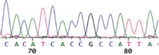
4.10.6 | In front of the primer name you should see another arrow. When this arrow is clicked the coloured trace is revealed.
|
5. Results
5.1 Sequencing results
5.1.1 | The results of this work should be reported to the project consultants or named signatories immediately upon completion. A second checker must check that you have correctly transcribed the sequence of the cleavage site. |
5.1.2 | The common cleavage site nucleotide sequences (and amino acid motifs), and points for their interpretation are listed in Appendix 1 and 2. Decide whether your isolate is HP or LP. |
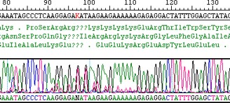
5.1.3 | Sequencing results: Fit for purpose Sequencing chromatograms which span the cleavage site region should be assessed critically, two examples are shown below. The top chromatogram represents a single sequencing run (ie in one orientation) where in places the quality of the data is poor (ie superimposed peaks of more than one signal, a general background pattern of red (ie T) signals), as seen from the character of the plot. This results in clearly one ambiguous nucleotide call at the cleavage site, and likely errors among neighbouring nucleotides which upon translation results in poor amino acid sequence data at the cleavage site. Such a result would be considered “unfit for purpose” unless there was a complementary sequencing run available (ie in the opposite orientation) of clearly superior quality.
|
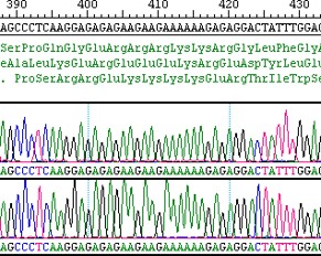
5.1.4 | The next example (below) consists of two complementary sequencing runs in opposite orientations. Compare the quality of the two below chromatograms with the above, ie clear and discrete peaks which agree in both orientations. There are no superimposed peaks nor is there any underlying background, and translation into amino acid sequence is perfectly reliable in giving a polybasic highly pathogenic AI cleavage site in this example. Clearly the below data is “fit for purpose”.
|
5.2 Troubleshooting
5.2.1 | This PCR relies on the quality of the RNA template being good. If you fail to get an electrophoresis band you should obtain a new extraction of RNA and repeat the test. |
5.2.2 | In most instances the primers mentioned in section 4.4 and 4.5 will produce a good PCR product and you will see a clear band of the expected size on the electrophoresis gel. If these primers do not produce a good band it may be necessary to try some alternatives. Be sure to record which primers you used in the appropriate Statutory sequencing book. |
5.2.3 | Alternative sequencing primers can also be used when necessary. |
6. References
6.1 | Slomka MJ, Coward VJ, Banks J, Löndt BZ, Brown IH, Voermans J, Koch G, Handberg KJ, Jørgensen PH, Cherbonnel-Pansart M, Jestin V, Cattoli G, Capua I, Ejdersund A, Thorén P and Czifra G (2007). Identification of sensitive and specific avian influenza polymerase chain reaction methods through blind ring trials organized in the European Union. Avian Diseases, 51: 227-234 |
Appendix 1 – Interpretation of H5 cleavage site results
An avian influenza virus is considered HPAI if there are multiple basic (arginine - R or lysine- K) amino acids at the HA cleavage site. Most HPAI virus HAs have the sequence motif R-X-R/K-R-G (X can be any amino acid) although there are exceptions. In practice any motif with more than two basic amino acids should be considered HPAI or to be at risk of becoming HPAI. The cleavage site usually begins with a proline - P and ends with RGLF.
Examples of H5 AI amino acid sequences across the cleavage site (not inclusive)
Virus | Subtype | Motif | Pathogenicity1 |
Numerous LPAI Eurasian viruses | H5N* | PQRET-------- RGLF | - |
A/turkey/Kfar Vitkin/71 | H5N2 | PQREA-------- RGLF | - |
A/turkey/Ramon/73 | H5N2 | PQREA-------- RGLF | - |
A/laughing gull/AK/296/75 | H5N3 | PSIGE-------- RGLF | - |
A/arctic tern/AK/300/75 | H5N3 | PSIGE-------- RGLF | - |
A/mallard/Alberta/271/88 | H5N3 | PEKQT-------- RGLF | - |
A/chicken/Italy/RA9097/98 | H5N9 | PQKET-------- RGLF | - |
A/chicken/Scotland/59 | H5N1 | PQRKK-------- RGLF | + |
A/tern/South Africa/61 | H5N1 | PQRETRRQKRGLF | + |
A/turkey/Ontario/7732/66 | H5N9 | PQRRKK---RGLF | + |
A/chicken/PA/1370/83 | H5N2 | PQKKK-------- RGLF | + |
A/duck/Ireland/113/83 | H5N8 | PQRKRKK--RGLF | + |
A/turkey/Ireland/1378/83 | H5N8 | PQRKRKK--RGLF | + |
A/turkey/England/91 | H5N1 | PQRKRKT--RGLF | + |
A/chicken/Pueblo/94 | H5N2 | PQRKRKT--RGLF | + |
A/chicken/Queretaro/20/95 | H5N2 | PQRKRKRKTRGLF | + |
A/chicken/Puebla/8623-607/97 | H5N2 | PQRRKRKTRGLF PQRKRKRKTRGLF | + |
A/chicken/Hong Kong/990/97, A/chicken/Thailand/04 and other clade 2-like H5N1 viruses | H5N1 |
PQRERRRKKRGLF | + |
A/Hong Kong/156/97 | H5N1 | PQRETRRKKRGLF | + |
A/Hong Kong/486/97 | H5N1 | PQRRRRKK-RGLF | + |
A/poultry/Italy/97 | H5N2 | PQRRRKK-RGLF | + |
A/chicken/Italy/1487/97 | H5N2 | PQRRKKR-RGLF | + |
A/avian/Texas/04 | H5N2 | PQRKKRGLF | + |
A/ostrich/ZA/N227/04 | H5N2 | PQREKRRKKRGLF | + |
A/ostrich/SA/AI2114/11 A/ostrich/SA/AI2887/11 | H5N2 | PQRRKKRGLF PQRRRKRGLF | + |
A/turkey/Turkey/1/05 and other clade 2.2 H5N1 viruses | H5N1 | PQGERRRKKRGLF | + |
Virus | Subtype | Motif | Pathogenicity1 |
A/chicken/Vietnam/19640/09 (clade 2.3.4) (and clade 2.3.4.4 H5N8 HP outbreaks in the EU and UK 2016-2017) | H5N8 |
|
|
Seen in some clade 2.3.4 H5N1 viruses 2009-2013 and clade 2.3.4.4 H5N8s | H5N1 H5N8 |
PLRERRRKRGLF | + |
H5N8 HP UK wild bird 2017 | H5N8 | PLRKKRRKRGLF | + |
Seen in some clade 1 H5N1 viruses 2009-2013 | H5N1 | PQREGRRKKRGLF | + |
Seen in some clade 1 H5N1 viruses 2009-2016 | H5N1 | PQREERRKKRGLF | + |
Seen in some clade 2.1 H5N1 viruses 2009-2016 | H5N1 | PQRESRRKKGLF | + |
Seen in some clade 2.3.1, 2.3.2 and 2.3.3 H5N1 viruses 2009-2013 | H5N1 | PQRERRRKRGLF | + |
A/duck/Vietnam/18151/09 (clade 2.3.2) | H5N1 | PQRERRRRKRGLF | + |
Seen in some clade 7 H5N1 viruses 2009-2013 | H5N1 | PQIEGRRRKRGLF | + |
A/chicken/Israel/93/2015 (clade 2.2.1) | H5N1 | PQGEKRRKKRGLF | + |
Seen in some clade 2.2 H5N1 viruses 2009-2016 | H5N1 | PQGEGRRKKRGLF | + |
A/chicken/Iraq/2016 | H5N1 | PQREKRRKRGLF | + |
A/chicken/France/150169a/2015 A/duck/France/150233b/2015 A/duck/France/150236b/2015 | H5N1 H5N2 H5N9 |
HQRRKRGLF | + |
Seen in some clade 4 H5N1 viruses 2000-2008 | H5N1 | PQREIRRKKRGLF | + |
1= Pathogenicity of viruses as determined by IVPI or equivalent tests. + = HPAI, - = LPAI
Appendix 2 – Interpretation of H7 cleavage site results
Examples of H7 AI amino acid sequences across the cleavage site (not inclusive)
Virus | Subtype | Motif | Pathogenicity1 |
Numerous American viruses | H7N* | PENPK--- TRGLF | - |
Numerous American and Eurasian | H7N* | PEIPK--- GRGLF | - |
A/ruddy turnstone/NJ/65/85 | H7N3 | PEKPK--- TRGLF | - |
A/duck/OH/10/88 | H7N8 | PESPK--- TRGLF | - |
A/finch/CA/28710-8/93 | H7N8 | PEIPK--- ERGLF | - |
A/pekin robin/CA/30412-5/94 | H7N1 | PEIPK---RRRGLF | - |
A/FPV/Brescia/02 | H7N1 | PSKKR---KKRGLF | + |
A/FPV/Dobson/27 | H7N7 | PELPK-KRRKRGLF | + |
A/FPV/Dutch/27 | H7N7 | PPKKR---RKRGLF | + |
A/FPV/Rostock/34 | H7N1 | PEPSK-KREKRGLF | + |
A/FPV/Weybridge | H7N7 | PELPK-KRRKRGLF | + |
A/FPV/Egypt/45 | H7N7 | FSKKR---RKRGLF | + |
A/turkey/England/63 | H7N3 | PETPK--RRRRGLF | + |
A/chicken/Victoria/76 | H7N7 | PEIPK-KKEKRGLF | + |
A/duck/Victoria/76 | H7N7 | PEIPK--- KRGLF | - |
A/turkey/England/199/79 | H7N7 | PEIPK-KREKRGLF | + |
A/chicken/Leipzig/79 | H7N7 | PEIPK--KKGRGLF | + |
A/goose/Leipzig/192/79 | H7N7 | PEIPKRKKRGLF PEIPKKRKKRGLF PEIPKKKKKKRGLF | + |
A/chicken/Victoria/1/85 | H7N7 | PEIPK-KREKRGLF | + |
A/chicken/Queensland/94 | H7N3 | PEIPRKRKRGLF | + |
A/chicken/Pakistan/447/94 | H7N3 | PETPK-RKRKRGLF | + |
A/chicken/Pakistan/547-99/95 | H7N3 | PETPK--RRNRGLF | + |
A/chicken/Pakistan/1369/95 | H7N3 | PETPK--RRKRGLF | + |
A/chicken/New South Wales/2/97 | H7N4 | PEIPRRRKRGLF | + |
A/Teal/Taiwan/19. 2-37-2/98 | H7N1 | PEIPK-GRGLF | - |
A/poultry/Italy/1999 | H7N1 | PEIPKGSRVRRGLF PEIPKGSRMRRGLF PEIPKRSRVRRGLF | + |
Italy/7159-V02/02 | H7N3 | PEIPKGRGLF | - |
Pakistan/various/01 | H7N3 | PETPK--RRKRGLF | + |
Pakistan/27/01 | H7N3 | PEIPKGRGLF | - |
Pakistan/33/ 01 | H7N3 | PETPKRRKRGLF | + |
Virus | Subtype | Motif | Pathogenicity1 |
A/poultry/Chile/2002 | H7N3 | PEKPKTCSPLSRCRETRG LF PEKPKTCSPLSRCRKTRG LF | + |
A/poultry/Chile/2002 | H7N3 | PEKPKTRGLF | - |
A/chicken/NL/2003 | H7N7 | PEIPKRRRRGLF | + |
A/turkey/NL/2003 | H7N3 | PEIPKGRGLF PEIPKGSRVRRGLF | - + |
A/ck/Pakistan/03 | H7N3 | PETPK-RRKRGLF | + |
A/chicken/Canada/rv504/04 | H7N3 | PENPKQAYRKRMTRGLF PENPKQAYQKRMTRGLF PENPKQAYKKRMTRGLF PENPKQAYHKRMTRGLF PENPKQAHQKRMTRGLF PENPRQAYRKRMTRGLF PENPKQACQKRMTRGLF | + |
A/chicken/North Korea/1/2005 | H7N7 | PEIPKGRHRRPKRGLF | + |
A/turkey/Italy/4527/2007 | H7N3 | PETPKGRGLF | - |
A/chicken/Saskatchewan/HR-00011/07 | H7N3 | PENPKTTKPRPRRGLF | + |
A/mallard/Sweden/100993/08 | H7N7 | PEIPKKRGLF | - |
A/chicken/England/2008 | H7N7 | PEIPKRKKRGLF PEIPKKKKRGLF PEIPKKKKKKRGLF | + |
A/goose/Czech/NRL 1848/09 | H7N9 | PELPKGRGLF | - |
A/chicken/Spain/6279-2/09 | H7N7 | PELPKGTKPRPRRGLF | + |
A/chicken/Italy/11VIR-62/11 | H7N3 | PEPPKGRGLF | - |
A/chicken/Italy/13VIR4527-11/13 | H7N7 | PETPKRRERRGLF | + |
A/chicken/England/2015 | H7N7 | PEIPRHRKGRGLF PEIPRHRKRRGLF | + + |
A/chicken/Germany/AR1385/2015 | H7N7 | PEIPKRKRRGLF | + |
A/chicken/Italy/16VIR-1873/2016 | H7N7 | PELPKGRKRRGLF | + |
A/turkey/Indiana/16-001403-1/2016 | H7N8 | PENPKKRKTRGLF | + |
A/chicken/Jalisco/CPA1/12 A/chicken/Puebla/CPA-04451/16 A/chicken/Jalisco/CPA-01859/16 | H7N3 | PENPKDRKSRHRRTRGLF PENPKDRKNRHRRTRGLF PENPKGKKSRHRKTRGLF | + |
H7N9 2017 China and Taiwan human and chicken | H7N9 | PEVPKRKRTARGLF PEVPKGKRTARGLF | + |
1= Pathogenicity of viruses as determined by IVPI or equivalent tests. + = HPAI, - = LPAI